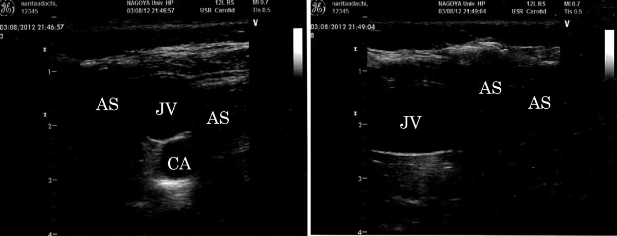
Figure 2

Ultrasound images. Left: The ultrasound image of the patient's neck in coronal view was demonstrated. The identification of both jugular vein and carotid artery was barely possible in spite of many subcutaneous ultrasound barriers by emphysema. Right: The ultrasound image in sagittal view was demonstrated. The jugular vein was feasibly observed; however, the carotid artery that is a relatively deep structure could not be identified. JV jugular vein, CA carotid artery, AS acoustic shadow by air.
